Diphtheria – Epidemiology, Clinical Features & Management
Introduction:
- Three major clinical types
- Anterior nasal
- Faucial and
- Laryngeal
- Rarely, other body parts may be affected
- Skin
- Conjunctiva
- Vulva etc.
- The bacilli multiply locally (mostly this site is the throat) and elaborate a powerful exotoxin which causes the following:
- Formation of a greyish or yellowish membrane commonly over the
- tonsils,
- pharynx or
- Larynx or
- At the site of implantation
- This is a false membrane with
- Well defined edges and
- Cannot be wiped away
- Marked congestion, edema or local tissue destruction
- Enlargement of regional lymph nodes
- Signs and symptoms of toxemia
- Average Fatality rate is about 10% in untreated cases
- Fatality rate in under 5 yr. children is 20% of untreated cases
Problem Statement
- World
- Rare disease in the developed countries
- Reason is routine vaccination of children
- Countries with satisfactory vaccination schemes,
- the disease has declined to a level that it is no longer a PHP
- A case may occasionally be seen among the non immunized children in developed countries
- Natural immunity against diphtheria is acquired by frequent skin infection with C. diphtheriae
- Improved SE conditions have changed this situation in developed world leading to a change in epidemiology of diphtheria
- Some outbreaks have demonstrated a shift in the age distribution of cases to older children and adults
- In developing countries
- The disease is endemic due to lack of widespread immunization
- India
- Diphtheria is endemic
- Declining trend, due to increasing immunization coverage
- CFR of about 2.6%
Agent Factors
- Agent is C diphtheriae
- Gram positive
- Non motile bacilli
- No invasive power
- Produces a powerful exotoxin
- Four types of bacilli, all are pathogenic to man
- Gravis
- Mitis
- Belfanti and
- Intermedius
- In general, gravis infections tend to be more severe than mitis infection
- All strains are not toxigenic
- Evidence suggests that non–toxicogenic strain can convert to toxicogenic upon exposure to the beta – phage (a bacteriophage)
- The toxin can affect
- The heart, leading to myocarditis or
- The nerves, leading to paralysis
- Sensitivity:
- Sensitive to Penicillin
- Sensitive to
- Heat and
- chemical agents
- They survive for short periods in dust and fomites
Source of infection
- Case or
- Range from subclinical to frank clinical
- Mild to moderate cases may present as only running nose or sore throat
- These play more important role than frank cases in the spread of infection
- Carrier
- Common source of infection; Carrier: cases = 95: 5
- Carrier may be
- Temporary or chronic
- Nasal or throat
- Nasal carriers are shed more frequently and hence more dangerous as source
- Temporary, carrier state may last about a month; chronic carrier state may persist about a year, unless treated
- Incidence of carriers in a community varies 0.1 – 5%
- Immunization does not prevent carrier state
Infective material
- Nasopharyngeal secretions
- Discharge from skin lesions
- Contaminated fomites and
- Possibly infected dust
Period of infectivity
- Unless treated, lasts from 14 days to 28 days from the onset of the disease
- Carriers remain infective for much longer periods
- A case or carrier is considered non – communicable:
Host Factors
Age
- Affects particularly children aged 1 yr. to 5 yr.
- In countries with widespread immunization, a shift in age incidence has been observed from preschool to school age
Sex:
- Both sexes are affected
Immunity
- Infants born of immune mothers are relatively immune during first few weeks or months of life
- Before artificial immunization,
- large proportion of population in developing countries were acquiring active immunity through in - apparent infection resulting in widespread production of antitoxin in the population
- Thus most members of the population except children were immune
- By age 6-8 yr. appx 75% of children in developing countries where skin infection with C are common, have protective serum antitoxin levels
- Diphtheria is principally the result of action of the toxin formed by the organism rather than invasion by the organism,
- resistance to the disease depends largely on the availability of specific neutralizing antitoxin in the bloodstream and tissues
- Diphtheria occurs only in persons who possess no antitoxin
Environmental factors
- Cases occur all seasons
- Winter months favor its spread
Mode of transmission
- Mainly by droplet infection
- Can also be transmitted directly to susceptible persons from infected cutaneous lesions
- Transmission by contaminated objects like cups, thermometers, toys, pencils is possible but only for short periods
Portal of entry:
- Respiratory route
- Commonly the portal of entry
- Non – respiratory route:
- Skin: cuts, wounds and ulcers may get infected
- Umbilicus in the newborn
- Occasional ports of entry
- Eye
- Genitalia or
- Middle ear
- Less common in developed countries, here spread by droplet infection is more common
Clinical Features
- IP: 2 to 6 days, occasionally longer
- Respiratory tract forms:
- Pharygotonsillar
- Laryngotracheal
- Nasal and
- Combinations
Pharyngotonsillar diphtheria
- Sore throat, difficulty in swallowing and low grade fever
O/E:
- Mild erythema throat
- Localized exudate or pseudo-membrane
- On posterior wall of pharynx or on tonsil
- May cover entire tonsil or less frequently even the soft and hard palates
- Pseudo membrane may be whitish and wipe off easily in early stage
- Later the membrane may become thick, blue-white to grey-black and adherent
- Attempts to remove the membrane result in bleeding
- A minimal area of mucosal erythema surrounds the membrane
- In severe disease
- Marked edema of submandibular area and anterior neck and
- Lymphadenopathy giving a characteristic ‘bull necked appearance
Laryngotracheal diphtheria
- Often preceded by pharyngotonsillar disease
- Fever
- Hoarseness
- Croupy cough
- If infection extends into bronchial tree, it is the most severe form of disease
- Initially the may be indistinguishable from viral croup or epiglottitis
- Prostration and dyspnea soon follow because of the obstruction by the membrane
- This obstruction may even cause suffocation if not promptly relieved by intubation or tracheostomy
Nasal Diphtheria
- Mildest form
- Localized to the septum or turbinates of one side of the nose
- Occasionally the membrane may extend into the pharynx
Cutaneous diphtheria
- Common in tropical area
- Often appears as a secondary infection of a previous skin abrasion
- Presenting lesion is often an ulcer
- Ulcer is
- Surrounded by erythema
- Covered with a membrane
- Patients generally seek treatment because of the chronicity of the skin lesion
- The bacilli within the membrane continue to produce toxin actively
- This is absorbed and results in distant toxic damage, particularly
- parenchymatous degeneration, Fatty infiltration and necrosis in
- Heart muscle
- Liver
- Kidneys and
- Adrenals
- Irregularities in cardiac rhythm indicate damage to the heart
- Later there may be difficulties with
- Vision
- Speech
- Swallowing or
- Movement of the arms or legs
- The toxin also produced nerve damage, leading to paralysis of the
- Soft palate
- Eye muscles or
- Extremities
- Those who survive these complications, recover completely
Control of Diphtheria
- Treatment of
- Cases and
- Carriers
- Management of the ‘Contacts’ and
- At the community level
- Increasing coverage of immunization with diphtheria toxoid
Treatment of Cases and Carriers
Early detection
- An active search for cases and carriers should start immediately amongst family and school contacts
- Carriers can be detected ONLY by culture
- Swabs should be taken from both the nose and throat
- Examined by culture for diphtheria bacilli
- Tests should also be done for virulence of the organism
Isolation
- All cases
- Suspected cases and
- Carriers should be
- Promptly isolated
- Preferably in a hospital
- For at least 14 days or
- Until proved free of infection
- At least 2 consecutive
- nose and throat swabs,
- taken 24 hours apart
- should be negative before terminating isolation
Treatment of cases and carriers
- Cases:
- Diphtheria antitoxin should be given without delay,
- IM or IV, Dose ranges from 20,000 to 1,00.000 units depending up on the severity
- Mild early pharyngeal or laryngeal disease: 20 to 40,000 units
- Moderate nasopharyngeal disease : 40 to 60, 000 units
- For severe, extensive or late (≥3 days): 80 to 1,00,000 units
- Penicillin or erythromycin for 5 – 6 days to clear the throat of C. diphtheriae and thereby decrease toxin production
- The case should be reported to the health authorities Immediately
- Once recovered, active immunization with diphtheria toxoid should be provided
- Carriers:
- Treat with 10 days course of oral erythromycin
Management of Contacts
Contacts merit special attention
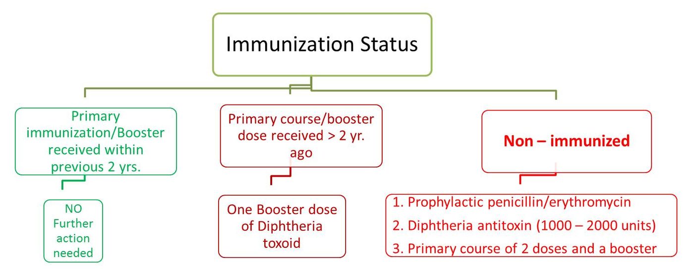
- They should be throat swabbed
- Determine the immunization status and take further action accordingly (shown below)
- Contacts need to be placed under medical surveillance
- Examined daily for evidence of diphtheria for one week
- Bacteriological surveillance weekly for several weeks (weekly swabbing)
References
- DIPHTHERIA - A RE-EMERGING THREAT. CD Alert, Jan – Mar 2008; 12 (1): 1-8 available on http://www.ncdc.gov.in/writereaddata/linkimages/Jan-March%20087398246488... downloaded on 16 Dec 2016
- Communicable Disease Management Protocol – Diphtheria. August 2016. available at: http://www.gov.mb.ca/health/publichealth/cdc/protocol/diphtheria.pdf downloaded on 16 Dec 2016
- http://www.immunize.org/catg.d/p4203.pdf accessed on 16 Dec 2016
- Park’s Textbook of Preventive and Social Medicine, 23rd Edition, Diphtheria, 161-163
enable_page_level_ads: true
});
Control of diphtheria: Control of Diphtheria
Management of Contacts of a Case of Diphtheria: Management of Contacts of a Diphtheria Case
Diphtheria – Epidemiology, Clinical Features & Management: Diphtheria – Epidemiology, Clinical Features & Management