Arterial Pulse: Clinical Examination and Physiology
1. The Physiology of the Arterial Pulse
In an era increasingly dominated by high-tech diagnostic imaging, the arterial pulse remains a dynamic window into the cardiovascular system. It serves as a vital clinical bridge, allowing the clinician to translate complex cardiac physiology into actionable bedside data.
Mastering its assessment is not merely about counting beats; it is about interpreting the mechanical output of the heart in real-time.
The Pressure Wave Mechanism
The arterial pulse is generated by the sudden, forceful ejection of blood from the left ventricle into the arterial network. This ejection causes the elastic walls of the arteries to distend.
Clinically, we define the arterial pulse as the pressure impulse felt by the fingertips when a superficial artery is pressed against an underlying firm structure, such as a bone.
This sensation is the physical manifestation of the pressure wave expanding the arterial wall as it travels from the heart to the periphery.
The Systolic Correlation
The peak of this transmitted pulse wave correlates closely with the patient's systolic blood pressure.
By evaluating the strength and timing of this peak, we gain immediate insight into the efficacy of ventricular contraction and the state of the systemic circulation.
Understanding this physiological "how"—the interplay between ventricular ejection and arterial elasticity—is the necessary precursor to mastering the "how-to" of the physical examination.
2. Step-by-Step Radial Pulse Examination Technique
While many sites are available for palpation, the radial artery at the wrist is the primary site for initial clinical assessment.
The patient’s forearm should be semi-pronated and the wrist slightly flexed.
This posture relaxes the surrounding tendons and allows the clinician to effectively press the radial artery against the underlying radius bone.
The Three-Finger Technique
The examination is performed using the index, middle, and ring fingers of your hand to palpate the patient's right radial pulse.
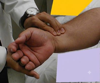
Apply pressure with these three fingers over the radial artery until the maximum impulse is felt. Using three fingers provides a larger surface area for detection and allows for a more nuanced assessment of the wave's character.
Senior Faculty Tip: Beginners often confuse the patient’s pulse with their own digital pulse; If you are uncertain, verify by feeling your own superficial temporal pulse on the side of your forehead with your other hand and compare the rhythms.
* The Training Phase: During the initial training period, always count the pulse for a full 60 seconds to develop a "feel" for the rhythm and rate.
* The Experienced Shortcut: Once sufficient experience is gained, you may count for 30 seconds and multiply by 2, or 15 seconds and multiply by 4.
* The 1-Minute Rule: Regardless of experience level, if the pulse is too slow, too fast, or irregular, you must count for a full 60 seconds to ensure an accurate reading.
3. The 8 Pulse Features to Evaluate
To transform a simple palpation into a diagnostic tool, the clinician must utilize a systematic framework. The examination usually includes a review of the following eight features:
1. Rate: The normal resting range for an adult is 60–100 beats per minute (bpm).
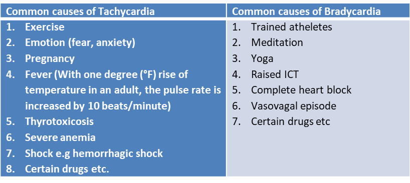
Tachycardia is defined as >100 bpm, and bradycardia is <60 bpm.
Notably, for every 1°F increase in body temperature, the pulse rate typically increases by 10 bpm.
2. Rhythm: Determine if the pattern is regular, regularly irregular, or irregularly irregular.
3. Volume (Amplitude): This reflects pulse pressure and left ventricular stroke volume. While felt at the radial, it is best evaluated at the carotid artery.
4. Character (Waveform): This involves the contour of the wave (upstroke, peak, and downstroke) and is also best assessed at a central artery e.g. the Carotid artery.
5. Radio-radial Delay: Palpate both radial pulses simultaneously. A discrepancy in timing or volume may indicate atherosclerosis, subclavian artery stenosis, or an aortic dissection.
Note that atheromatous narrowing of the axillary artery can specifically reduce pulse strength on the affected side.
6. Radio-femoral Delay: Palpate the radial and femoral pulses simultaneously.
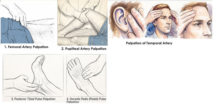
To palpate the femoral pulse, press deeply below the inguinal ligament, midway between the anterior superior iliac spine (ASIS) and the symphysis pubis, feeling the pulse against the femur.
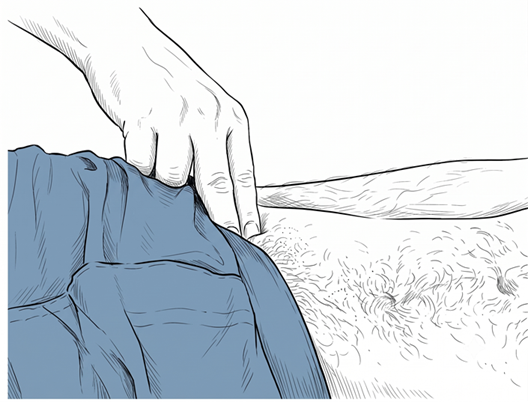
A perceptible delay suggests Coarctation of the Aorta (a narrowing of the aortic isthmus), which is often associated with upper limb hypertension.
7. Vessel Wall Condition: Roll the relaxed radial artery against the radius. In elderly patients, thickening or tortuosity may be felt; however, this is a general observation and does not always indicate a narrowed lumen.
8. Peripheral Pulses: A complete survey must include all palpable peripheral sites to ensure systemic vascular health.
Clinical Drivers of Rate
The following table summarizes the physiological and pathological factors influencing pulse rate:
4. Deciphering Irregular Rhythms and the Pulse Deficit
Ascertain if the rhythm of the pulse is regular or irregular.
Regular rhythm: the time between beats remains constant
Irregular rhythms: when the time between beats is varying.
Irregular rhythm may be: Regularly irregular OR Irregularly irregular
Identifying an "irregular" pulse is of high diagnostic value, as it often signals underlying electrical or mechanical instability.
Regularly Irregular Patterns
In these cases, the irregularity follows a predictable recurring pattern:
* Sinus Arrhythmia: A common and normal finding where the pulse rate increases during inspiration and decreases during expiration.
* Ectopic Beats: Extra beats occurring in fixed ratios. Bigeminy occurs when every second beat is ectopic (normal pulse, weak pulse, delay). Trigeminy occurs when every third beat is ectopic.
* Wenckebach Phenomenon: A pattern where AV node conduction time increases progressively until an atrial systole is completely dropped. The cycle then resets, creating a pulse that feels like clustered beats followed by a pause. The pulse feels like a repeating pattern of clustered beats followed by a pause
E.g. tap... tap... tap... [pause]... tap... tap... tap... [pause]…….
Irregularly Irregular Patterns: This represents a chaotic rhythm with no discernible pattern.
* Atrial Fibrillation (AFib): The SA node fires at rates potentially >600/min.
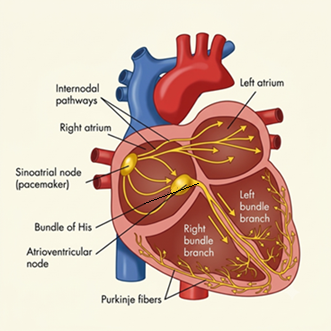
The AV node protects the ventricles by blocking a variable proportion of these impulses. The resulting heart rate is often fast (150–180/min) and entirely chaotic, with varying pulse volumes due to variable diastolic filling times.
Other causes include frequent irregular ectopic beats and Atrial Flutter with variable block.
The Pulse Deficit Protocol
When a rhythm is irregular, the clinician should check for a Pulse Deficit using a two-person technique: one person listens to the heart with a stethoscope while the other simultaneously palpates the radial pulse.
* The Finding: A pulse deficit is present if the auscultated heart rate is higher than the palpable radial pulse rate.
* The Significance: This occurs because a disturbed rhythm may result in inadequate ventricular filling time. The resulting contraction lacks the stroke volume necessary to produce a palpable peripheral wave.
Essentially, the heart beat occurred, but the peripheral pulse did not.
5. Clinical Pulse Volume and Waveform Contours
Assessing the "Character" of the pulse is the most advanced skill in palpation, requiring a deep understanding of hemodynamics.
Assessment Sites
Central arteries (Carotid or Brachial) are superior for volume and character because their proximity to the left ventricle prevents the distortion of the pressure wave found in more distant vessels.
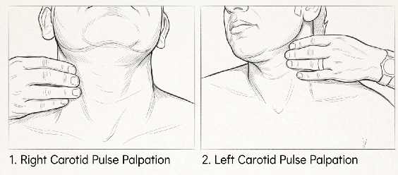
When palpating the carotid, place the patient at a 30° angle. Use the pulp of your fingers or thumb medial to the sternocleidomastoid muscle.
Gradually increase pressure until you feel a maximal pulsation; then slowly decrease pressure until you best feel the arterial pressure and contour
Apply light pressure to compress the artery partially, allowing you to gauge the ‘Volume’ (force of the impulse) and ‘Character’ (contour).
For the left carotid artery, use your right fingers or thumb
Safety Note
• Never palpate both carotid arteries simultaneously to prevent carotid sinus syncope or cerebral hypo-perfusion
• Be careful to avoid pressing the carotid sinus, which lies adjacent to the top of the thyroid cartilage
- Pressure on the carotid sinus may cause reflex bradycardia or drop in blood pressure.
Pulse character describes the waveform contour felt in a single pulse wave
- The rate of upstroke (anacrotic limb),
- The contour of the peak, and
- The rate of down-stroke (dicrotic limb)
Focus on the contour of the pulse wave: Try to feel: - How fast it rises to a peak,
- How long the peak is sustained, and
- How rapidly it collapses.
The normal - Upstroke is brisk; smooth and follows S1 almost immediately.
- Summit is smooth, rounded, and around midsystolic.
- Down-stroke is less abrupt than the upstroke
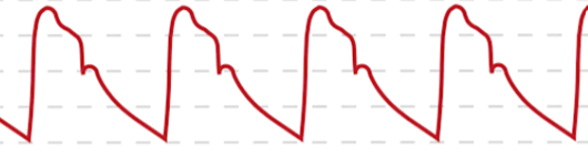
Pathological Waveform Reference
A sophisticated examiner looks for specific contours in the pulse wave:
Pulsus Parvus et Tardus
* Description: Small amplitude (parvus) with a delayed, slow systolic upstroke (tardus).
* Mechanism: Fixed obstruction to left ventricular outflow delays peak pressure delivery.
* Clinical Significance: Severe Aortic Stenosis.
Collapsing / Water-Hammer Pulse
* Description: A rapid upstroke followed by a sudden collapse.
* Technique: Best felt at the radial artery. Use the flat palmar surface of your fingers and swiftly elevate the patient’s arm vertically above their head to accentuate the collapse.
* Clinical Significance: Aortic Regurgitation or high-output states.
Bisferiens Pulse
* Description: A double systolic peak felt within a single cardiac cycle.
* Mechanism: A rapid initial ejection peak followed by a secondary tidal wave.
* Clinical Significance: Combined AS + AR or HOCM.
Pulsus Alternans
* Description: A regular rhythm with beat-to-beat alternation in volume (strong beat, weak beat).
* Mechanism: Alternating strength of myocardial contraction due to diseased myocytes.
* Clinical Significance: Advanced Heart Failure.
Pulsus Paradoxus
* Description: An exaggerated reduction in pulse volume during inspiration.
* Mechanism: Rigid pericardial restraint causes the septum to bulge, compromising LV filling during inspiration.
* Clinical Significance: Cardiac Tamponade, Constrictive Pericarditis, or severe Asthma.
Dicrotic Pulse
* Description: A double peak with the second peak occurring in early diastole.
* Mechanism: Low cardiac output combined with low systemic vascular resistance.
* Clinical Significance: Sepsis or severe dilated cardiomyopathy.
Palpate the Peripheral Pulses
A complete clinical examination requires the survey of multiple peripheral sites to ensure systemic perfusion is adequate.
* Popliteal: Either flex the knee to 120° and use both hands in the fossa, or place the patient prone with the knee at 90°, resting the leg on your shoulder while pressing your thumbs deep into the fossa.
* Dorsalis Pedis: Palpate lateral to the Extensor Hallucis Longus (EHL) tendon. Press the artery against the tarsal bones. Note that it is absent in approximately 10% of healthy individuals.
* Posterior Tibial: Place the patient's foot relaxed, in the middle of plantar and dorsiflexion. Press behind and slightly below the medial malleolus of the ankle.
* Superficial Temporal: Palpate just in front of the tragus of the ear.
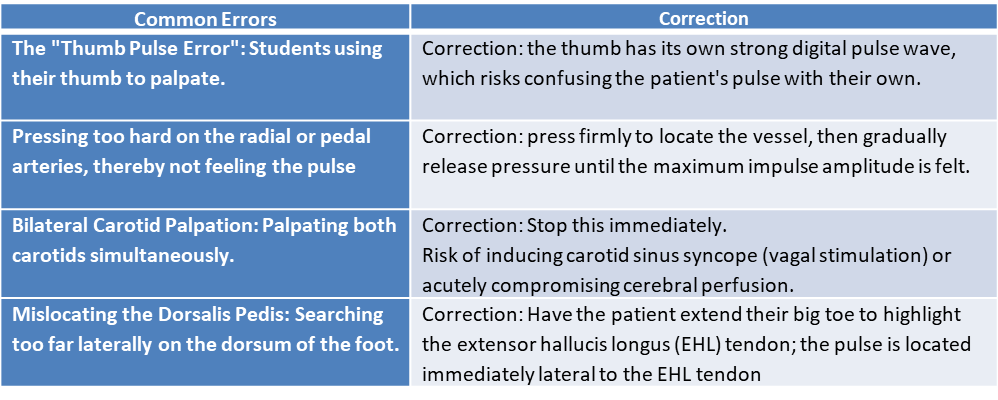
The Correction Corner
Final Summary
In the era of high-tech medicine, the arterial pulse remains one of the most cost-effective and immediate diagnostic tools available. It provides a real-time assessment of heart rhythm, valve function, and systemic vascular health.
As you continue to develop your clinical skills, remember the most accessible teacher is often yourself: check your own pulse regularly to master the nuances of the human heart.
References:
- Bickley, L. S., & Szilagyi, P. G. (2020). Bates' Guide to Physical Examination and History Taking (13th ed.). Wolters Kluwer.
- Michael Glynn, William M. Drake. Doctor and patient: General principles of history taking .In HUTCHISON’S CLINICAL METHODS. 24th Ed. London. Elsevier, 2018
- Kundu AK. Beginner's Guide to Bedside Medicine: Bedside Clinics in Medicine, Part I. 6th ed. New Delhi: Jaypee Brothers Medical Publishers; 2011.
- R Alagappan. In Manual of Practical Medicine 4th ed. New Delhi: Jaypee Brothers Medical Publishers; 2011
- Longo, D. L., Fauci, A. S., Kasper, D. L., Hauser, S. L., Jameson, J. L., & Loscalzo, J. (Eds.). (2022). Harrison's Principles of Internal Medicine (21st ed.). McGraw-Hill.
- Talley, N. J., & O'Connor, S. (2021). Clinical Examination: A Systematic Guide to Physical Diagnosis (9th ed.). Elsevier.
- National Center for Biotechnology Information (NCBI). Clinical Methods: The History, Physical, and Laboratory Examinations. Examination of the Extremities: Pulses, Bruits, and Phlebitis. Available via [National Library of Medicine Bookshelf](https://www.ncbi.nlm.nih.gov/books/NBK381/?utm_source=gemini).
Video for this blog: https://youtu.be/SAnhEHohmM4
Hindi video for this blog: https://youtu.be/UL-ttci5RVs